Scabies Infection – The Mite Bite

Photo credit:  http://www.skindermatologists.com/p/scabies-lice.html


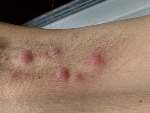
Every now and then a patient will come into the clinic with a scabies infection.  They usually have complaints of severe itching and a reddish colored rash.

Scabies is caused by a very small mite called Sarcoptes scabiei.  It is spread from one person to another by close skin-to-skin contact and I’m seeing more and more patients with scabies lately so I think it’s becoming more common.

Symptoms:  An itchy rash is the most common symptom, and it’s often worse at night.  It causes visible lesions (red colored bumps or blisters) on the skin but sometimes these little bumps are tough to see.  Certain parts of the body are most commonly affected by scabies including:

1)   Between the fingers

2)   Around the wrists (especially on the inside of the wrists)

3)   In the crease of the elbows

4)   Behind the knees

5)   In the armpits

6)   Around the nipples

7)   Around the penis

8)   Around the waistband

9)   Near the low back and upper thighs

10)  Along the sides and bottoms of the feet

The back and head are usually not affected.

Crusted scabies: Some people with weakened immune system can develop “crusted scabies” or “Norwegian scabies” which are described as large, crusty red patches or bumps on the skin that spread easily.  The scalp, hands, and feet are affected most often.  These lesions are usually not itchy but can contain many mites.

Scabies mite:   It is caused by a tiny mite that has 8 legs and is whitish-brown in color.  Without a magnifying glass, you might not be able to see them at all.  The symptoms are caused by the female mites, which tunnel into the skin after being fertilized by the males.  The female mite lays eggs under the skin and continues to tunnel until she dies, usually 1-2 months later.  After the mites hatch, the young mites travel back to the skin surface, mate and repeat the cycle of tunneling and laying more eggs.

Transmission:  Close skin-to skin-contact is the usual way that scabies is spread, but it can also be spread through the clothes of an infected patient.  If someone who is uninfected wears a shirt or jacket of someone who is infected, the little mites can infect another patient.  It takes about 3-4 weeks for signs or symptoms of a first scabies infection to develop after becoming infected with the mites.  It’s also commonly transmitted between young adults during sexual contacts.  Once the mites are no longer in contact with the skin, then can only live for 24-36 hours but they can survive longer in colder conditions.  They are seen more commonly in the winter than in the summer months.

Treatment:  Treatment of scabies can be challenging – see recommendations below.  Most of the time we treat scabies with a topical skin cream called permethrin (also called Elimite).  For patients with the more difficult to treat – crusted scabies, we use both a topical and oral anti-parasitic pill called ivermectin.  The permethrin cream (5%) is preferred for young infants and pregnant mothers.

1)  It is very important to apply the cream carefully to cover all the skin from the neck down to the feet.

2)  Treat all family members if they are in close contact with the infected person even if the family members don’t have symptoms.  The reason is to avoid repeating the cycle of infection.

3)  Wash or isolate any clothing, bedding, towels, pajamas, underwear or stuffed animals that the patient has touched within the last three days before the treatment started.  You can place the items in a plastic bag for three days to isolate them and the mites will die.  You can also wash the clothing in hot water.

Itching can be treated with antihistamines such as Claritin or Zyrtec.  Benadryl is helpful, but is sedating so we generally only recommend that at night.  Itching may persist for several weeks even after the mites are eliminated.  A steroid cream or a course of oral steroids may be recommended if itching is severe.

This document is for informational purposes only, and should not be considered medical advice for any individual patient.  If you have questions please contact your medical provider.


I hope that you have found this information useful.  Wishing you the best of health,

Scott Rennie, DO

Blog: https://doctorrennie.wordpress.com

All about Cold Sores (Oral Herpes)

shutterstock_64505920A patient came in this morning with a cold sore on her lip and was asking about treatment options.  She was wondering if this might be a sexually transmitted infection.  He’s never had a cold sore before that she can remember, but her boyfriend has genital herpes.

Cold sores are blisters that can be extremely painful.  They are usually near the lips or inside the mouth.  Herpes simplex virus is the cause of these blisters.  There are several types of herpes virus.  Type 1 usually causes cold sores while type 2 usually causes genital herpes that affects the penis or vaginal area.  A patient may have type 1 that occurs in the genital area or type 2 that affects the mouth but this is rare.  To answer this patient’s question about whether she could have got herpes on her lip from her boyfriend with genital herpes – the answer is “yes, it is possible.”

What are cold sores and how do I know if I have them?  These are actually also called fever blisters and are painful sores that usually occur near the mouth or lips.  The first time a patient gets cold sores is usually when they are a young child.  They can occur on the lips, mouth, nose or throat and usually form a small blister that pops and then forms a scab.  They are usually very sore and can also be associated with body aches or fever.

Sometimes people think they have a cold sore when instead they might have a canker  sore which is not caused from a virus.  The first time a patient has cold sores, the symptoms are usually more severe than when they get them in the future.  The initial attack can last 10-14 days and there are usually more ulcers than just one.  Patients complain of pain, burning, tingling or itching around their mouth sometimes even before the blister forms.

A medical provider can usually diagnose a cold sore, but they are so common that once you’ve seen them or have had them in the past, you can usually recognize them yourself.  If you have questions about a sore around your mouth however it’s always a good idea to have it checked by a medical provider.

How are cold sores spread?  The herpes virus that causes cold sores spreads easily from one person to the next usually by kissing or sharing a beverage container or eating utensil such as a spoon.  It can also be spread by people who have oral sex with someone who has genital herpes.   Once you have been infected with cold sores once, even after the sores go away the virus stays in your body in the nerve fibers under the skin.  More sores can come out any time and can be spread to other people.  Cold sores often re-occur when you become sick or your immune system is under stress.

Do I need to see a medical provider if I get a cold sore?  If you have severe pain, increasing redness or swelling around the mouth, nose or lips or trouble swallowing you should probably be seen by a medical provider.  Sometimes cold sores can become secondarily infected with a bacteria and cause a cellulitis or abscess that needs to be treated with antibiotics.  If a sore around the mouth is not going away, it’s also important to have it checked out to make sure that it’s not a cancer or something unexpected.

How do I get rid of cold sores?  The first time someone has cold sores the infection is usually worse and treatment is often recommended.  The virus can be treated with medication however there is no treatment to totally cure someone who has had cold sores or the herpes infection because the virus continues to exist in the nerve fingers under the skin even after the blisters go away.  If the sores come back after the initial infection, the symptoms are usually not as severe and usually go away within 8 days or less and there is usually less pain.

People with mild symptoms of cold sores often do not require treatment.  Patients often ask for medications to help reduce the duration of the cold sores or to treat the pain that accompanies the blisters.  I usually recommend ibuprofen or naproxen for pain relief due to the blisters.  There are various over the counter treatments which are helpful to some patients with cold sores including various creams or gels such as Abreva.  Abreva works by stopping the virus from entering into your cells and blocking the virus’ ability to replicate.  Patients also often get relief by using Orajel which is a topical numbing medicine that relieves that pain but does not make the viral infection go away any faster.

Oral prescription medication are sometimes prescribed for cold sores if they are severe.  Acyclovir, valacyclovir, and famciclovir are common oral antiviral medications that can be used for severe oral herpes infections.  Acyclovir seems to work the best for most people.  These antiviral therapies are usually most effective if started within the first 2-3 days of symptoms.  Some patients take chronic antiviral medications to prevent recurrent outbreaks of cold sores if:  recurrences are frequent or bothersome to the patient (ie. associated with frequent disfiguring lesions and pain) or for patients who have frequent serious systemic complications such as erythema multiforme, eczema herpeticum, or recurrent asceptic meningitis.

How can I prevent getting cold sores?  As I mentioned above, if you get cold sores frequently or they are associated with serious other conditions your doctor may prescribe a medication to take every day or periodically to prevent infections.  If you already have cold sores, avoid excessive sunlight as this has been shown to trigger cold sores to return.  Decreasing stress, getting enough sleep and staying healthy are some common sense ways of hopefully reducing your chances of developing recurrence of cold sores.  When you have a cold sore, do not kiss anyone or share silverware, glasses or cups, lip balm or razors.  Avoid oral sex when cold sores are present.

This document is for informational purposes only, and should not be considered medical advice for any individual patient.  If you have questions please contact your medical provider.


I hope that you have found this information useful.  Wishing you the best of health,

Scott Rennie, DO

Blog: https://doctorrennie.wordpress.com

What is Hidradenitis suppurativa?

shutterstock_148330382A patient came into the clinic today and said she had a couple lumps in her left armpit (axilla) that have been very painful and draining some clear liquid.  She told me that she had one before in her right arm pit and she had it treated by taking antibiotics and a doctor also performed an incision and drainage.

Hidrandenitis suppurativa (HS) is a condition in which there is red, swollen painful bumps in places where the skin rubs together.  The nodules can make it difficult to move because they are so painful.  They can also smell foul and drain pus or blood.  These bumps may go away on their own, but often stay for weeks to months and often come back.

Causes:  This is not caused by being unclean – it’s not an infectious disease. Often the area may become irritated by shaving in that area or a certain deodorant or antiperspirant.   You cannot spread this to anyone else as it’s not contagious.  It is generally a genetic condition that is more common in first-degree relatives that have the condition.

The bumps or nodules are usually located in the:

1)   Armpits (axilla)

2)   In the groin

3)   Under the breasts (in women)

4)   On the inner thighs

5)   Buttocks

6)   Around or near the anus

Often the skin hardens and scars around the painful nodules and some can form tunnels under the skin.

Treatment:  Possible treatment options include:

1)   Antibiotic liquids or gels that you put on the affected areas – these actually work to reduce inflammation rather than treat infection

2)   Antibiotic pills – to reduce inflammation

3)   Injections of steroid medications into the areas to bring down the inflammation

4)   Some women take hormone treatments to improve their condition

5)   Surgery

Things you can do to reduce your symptoms:

1)   If you are overweight, lose weight because this condition is more common or severe in people who are overweight

2)   Try to avoid activities that cause your skin to rub against itself

3)   Do not wear tight-fitting clothes

4)   For people with recurrent infections, the use of an antibiotic lotion such as clindamycin 1% applied to the area twice a day can help prevent recurrence.

5)   Show and wash the tender areas everyday gently.  Do not scrub with a washcloth, brush or loofah

6)   Avoid smoking

7)   Use antiperspirants rather than deodorants

8)   Avoid exposure to hot, humid environments as much as possible

This document is for informational purposes only, and should not be considered medical advice for any individual patient.  If you have questions please contact your medical provider.


I hope that you have found this information useful.  Wishing you the best of health,

Scott Rennie, DO

Blog: https://doctorrennie.wordpress.com

Tick Bites – Will I Get Lyme disease?

shutterstock_17720305shutterstock_148613042Last night a patient came into the Urgent Care with a tick on his belly.  He recently returned from a trip to New York State and he was concerned about the possibility of developing Lyme disease.  When I looked at the tick, I noticed that it was small and almost translucent.  It was attached to his skin but its head was still clearly visible and had not burrowed below the surface of the skin on his abdomen.

Tick bites are common this time of year.  They occur on humans as well as animals such as dogs.  Many different types of ticks in the United States, only some of which are capable of transmitting infections.  The risk of developing an infection such as Lyme disease after being bitten depends upon the geographic location, season of the year, type of tick and how long the tick was attached to the skin.

The risk of acquiring an infection from a tick is actually quite low.  In the case of my patient, the tick hadn’t even taken a blood meal – ie. it was not engorged with blood.  His risk of developing Lyme disease from that tick was absolutely 0%.  Ticks transmit infection only after they have attached and become engorged with blood.  Deer ticks that transmit Lyme disease must feed for more than 36 hours before transmission of the organism called Borrelia burgdorferi.

If you come in to see me after you’re bitten by a deer tick (the type that carries Lyme disease), I would generally advise one of two approaches:

1)   Observe the area and treat with antibiotics only if signs of infection develop

2)   Treat with antibiotics immediately as a preventative measure

The individual patient’s history, the type of tick and how long it was attached and the patient’s wishes will help determine which approach to take.

How to Remove a Tick:  Some patients come in to see me after they’ve already tried removing a tick and have been unsuccessful or partially successful.  I commonly see patients who come in after they’ve removed only part of the tick and the head and are concerned because the tick head is still buried below the skin.  Here is the technique that I use to remove a tick:

1)   Do not attempt to use a match, cigarette, nail polish, Vaseline, liquid soap or kerosene because it may just irritate the tick and cause it to inject the harmful organism into the wound

2)   Use fine tipped tweezers to grasp the tick as close to the skin as possible

3)   Pull back gently but firmly using even, steady pressure without jerking or twisting the tick

4)   After removing the tick, wash the skin and hands with warm soapy water

5)   If any part of the tick is still in the skin, they generally come out on their own.  I don’t recommend attempting to remove little pieces of the tick at home as this can cause skin trauma and scarring.

6)   Go see your medical provider if you are concerned about not being able to remove the tick.

Treatment:  The Infectious Diseases Society of America recommends treatment with antibiotics preventatively only in people who meet ALL the criteria below:

1)   The attached tick was identified as an adult or nymph deer tick

2)   The tick was attached for more than 36 hours based on how engorged the tick appears and the amount of time since outdoor exposure

3)   Antibiotic treatment can begin within 72 hours of tick removal

4)   The area where the tick bite occurred was in an area where the organism B. burgdorferi infection rate is greater than 20% – generally in parts of New England, parts of the mid-Atlantic states and parts of Minnesota and Wisconsin.

5)   The patient can take doxycycline – i.e. the patient is not pregnant or breastfeeding a young child or allergic to this antibiotic.

If all the criteria above are met, the treatment is a single dose of doxycycline 200mg for adults and 4mg/kg up to a maximum of 200mg for children older than 8 years of age.

Symptoms of Lyme disease:  What the area where the tick bite occurred and observe for expanding redness.  The rash that is associated with Lyme disease is called erythema migrams (EM).  This rash is a salmon color usually and typically expands over a few days or weeks and can reach up to 8 inches in diameter.  The center of the rash tends to become skin colored (clear) as the rash grows in size.  This gives the rash a sort of “bull’s eye” appearance.  The rash generally doesn’t cause any symptoms.

Other associated symptoms of Lyme disease could include:

1)  A few days to a month after the bite:  fatigue, malaise, lethargy, mild headache, mild neck stiffness, aches, joint pain and enlarged lymph nodes.

2)  Weeks to months after the bite:  Inflammation of the heart, heart rhythm problems, meningitis, encephalitis, severe joint pain, multiple areas of rash, eye pain/vision problems, liver disease, kidney disease.

This document is for informational purposes only, and should not be considered medical advice for any individual patient.  If you have questions please contact your medical provider.


I hope that you have found this information useful.  Wishing you the best of health,

Scott Rennie, DO
Blog: https://doctorrennie.wordpress.com

Allergic Antibiotic Drug Reactions – Am I Truly Allergic to Penicillin?

shutterstock_124906745shutterstock_46021174I had a patient who came in recently with his parents because he broke out in a rash all over his body after starting an antibiotic for an infection in his throat.  The parents were obviously concerned that he was allergic to the antibiotic and wanted to know what to do to help him.

Allergies to medications, especially to antibiotics such as penicillin are common, but it’s sometimes confusing to know whether or not it’s a true drug allergy, a skin rash from the infection or perhaps not an allergy at all.  Yesterday a patient told me that they are allergic to penicillin, and when I asked them what happens he takes it he said that he’s unsure – he just knows he allergic because his father was. I was alarmed that he thought he had an allergy to a potentially life saving medication only because a family member had long ago told him that they had an allergy to penicillin.

About 10% of patients report an allergy to penicillin when asked, however most people who believe they are allergic can take penicillin without a problem either because they were never truly allergic or because there allergy to penicillin has resolved over time. Only about 20% of people will be allergic to penicillin 10 years after the initial allergic reaction if they are not exposed to it again during this time period.

Definition:  Penicillin is a common antibiotic that is prescribed for strep throat, ear infections as well as pneumonia and many other infections.  It is part of a family of medications called beta lactams which include: Penicllin G, amoxicillin, ampicillin, oxacillin, cloxacillin, dicloxacillin, piperacillin, and nafcillin.  A patient who is allergic to one of these penicillin medications is presumed to be allergic to any of them in this group.

The Reaction:  It’s very important to tell your medical provider in as much detail as possible what the reaction is that happens if you take the medication rather than just listing it as an allergy.  If your medical provider writes down that you are allergic to an antibiotic and it’s not a true allergy, this might mean that the next time they get an infection, you get a less effective or more toxic antibiotic.

It’s important to distinguish between a true allergy and “adverse reactions.” Adverse reactions are unexpected reactions that occur after taking a medication which are common but not true allergies.  Some patients report an adverse reaction as an allergy because they don’t want to be given this medication in the future.  For example, I had a patient tell me that they could not take prednisone because they are allergic.  When I asked what happens if he takes it, he replied – “I don’t like the taste.”   Unfortunately there are a limited number of medications, and they are most often grouped into families.  If you are truly allergic to one medication in the family, this eliminates the possibility of taking any other medication in the same family.  An entire group of potentially helpful medications might have been withheld from this patient only because he doesn’t like the taste of one of them in this group.  Another example of a non-allergic adverse reaction is nausea and/or diarrhea.  By listing penicillin as an allergy because the patient gets nausea or diarrhea after taking might lead to antibiotic failure or resistance which can be costly and prolong illness.

It’s important to keep in mind that the care that you receive by your medical providers when you are ill might be negatively impacted by an improperly labeled allergy in your medical records.

Rashes:  There are different kinds of rashes that can occur after taking penicillin or other antibiotics.  Some rashes such as hives are raised, intensely itchy and they come and go over hours. Another type of rash is flat, blotchy and spread over days but do not change by the hour and are less likely to represent a dangerous allergy.  These rashes start after several days of treatment.  We call these rashes a drug induced exanthem.  Taking a photograph of your rash and bringing it to your doctor may be helpful if the rash changes.

True Allergic Reactions:  hives, angioedema (swelling of the face/lips), throat tightness, wheezing, coughing, trouble breathing from asthma type reactions are all important to distinguish from “adverse reactions” as I mentioned above.  When you list a medication allergy, make sure you describe which of these symptoms that the medication caused.  These types of reactions only occur in 1-5% of people.  It is important to tell your medical provider if you have had any of these symptoms because a past history indicates that the patient might develop a more severe infection such as anaphylaxis in the future if given the medication again.

Anaphylaxis:  This is a true medical emergency caused by a potentially life-threatening allergic reaction.  The symptoms involve the allergic reaction as well as low blood pressure, trouble breathing, abdominal pain, swelling of the throat or tongue and or diarrhea/vomiting.

Penicillin Allergy Testing:  Testing for a penicillin allergy might be important ifin people who have a suspected penicillin allergy and require it to treat a life-threatening condition for which no alternative antibiotic is appropriate.  It may also be appropriate for people who have frequent infections and have suspected allergies to many antibiotics, leaving few options for treatment.  About 90% of patients tested will not have a penicillin allergy either because they lost the allergy over time or were never allergic in the first place.  We do not routinely do allergy testing in the primary care or urgent care setting, it is done under the supervision of an allergist.

Cephalosporin Allergy:  Allergic reactions are less common than reactions to penicillin.  People with a penicillin allergy have a small risk of having an allergic reaction to cephalosporins.  Cephalosporins are a class of antibiotics closely related to penicillin.  Some of these medications include cephalexin, cefaclor, cefuroxime, cefadroxil, cepradine, cefprozil, loracarbef, ceftibuten, cefdinir, cefditoren, cefpodoxime, and cefixime.

Treatment:  For true allergic reactions stopping the medications as soon as possible is obviously important.  The following is an example of what I might do for a severe allergic reaction, but it may differ if you go a different facility or depending on the circumstances.

For mild urticaria:  Observation and consider diphenhydramine 25-50 mg PO/IM or 25mg IV

For severe urticara:  Diphenhydramine 25-50mg PO/IM or 25mg IV, Corticosteroids/Solumedrol 80-125mg IV,  IV fluids and/or epinephrine at a dose of 0.3mg 1:1000 IM (Epi-Pen).  If giving epinephrine, I usually have the patient transported to the hospital because they will need monitoring for rebound allergic reaction once the epinephrine wears off.

For Laryngeal Edema:  Give O2 by mask 6-10L

For Anaphylaxis-like reactions:  Suction as needed, elevate legs, O2 10L by mask, IV fluids (NS or LR), and Epinephrine 1:1000 0.3mg IM (Epi-Pen).  For bronchospasm, add Albuterol MDI 2-3 puffs, Antihistamine: Diphenhydramine 25-50mg IM or IV and Corticosteroids/Solumedrol 80-125mg IV and await transport to the hospital

For Hypotension:  Elevate legs, Oxygen by mask, use IV fluids, Epi-Pen and await transport to the hospital

Vagal Reaction:  Elevate legs, 02 by mask at 10L, IV fluids (NS/LR wide open)

For Angina:  02 by mask at 10L, IV fluids:  Administer slowly, Nitroglycerine 0.4mg sublingually; may repeat p5 min x 3 doses, Morphine 2mg IV and await transport to the hospital

For hypertension:  02 by mast at 10L, IV fluids:  Administer slowly, Nitroglycerine 0.4mg sublingually, may repeat q5 minutes x 3 doses and await hospital transport

For seizures: Suction/Protect Airway and monitor for obstruction by tongue.  O2 by mask if not vomiting.  If caused by hypotension, treat accordingly and if uncontrolled consider anticonvulsant such as diazepam and await hospital transport

For hypoglycemia:  O2 by mask at 10L, IV fluids D5W or glucose tablet

I recommend that patients who have known severe allergies to insect or bee stings carry an EpiPen with them.

If you’d like more information about allergic reactions to medications, check out the American Academy of Allergy Asthma & Immunology website:  http://www.aaaai.org/conditions-and-treatments/library/at-a-glance/medications-and-drug-allergic-reactions.aspx

This document is for informational purposes only, and should not be considered medical advice for any individual patient.  If you have questions please contact your medical provider.


I hope that you have found this information useful.  Wishing you the best of health,

Scott Rennie, DO

Blog: https://doctorrennie.wordpress.com

It’s Cloudy in Seattle, so why Worry about Sunburn?

shutterstock_5810650Recently I’ve been noticing patients are coming in with sunburns even though the weather has been cloudy.  They are often unaware that the sun is causing damage to their skin at the time, and come in later with severe sunburns.  Even on cloudy days, it is important to protect your skin because UV radiation can pass through the clouds and cause sunburns.  In addition, UV rays can be reflected off surfaces like sand, snow, cement and water.

Prevention:  Here are several ways to help prevent sunburn:

1)   Avoid sun exposure:  If you plan to be out in the sun during the day, keep in mind that the suns rays are strongest between 10:00am and 4:00pm in the continental U.S. and avoiding exposure during these hours decrease your risk of burning.  Wearing a topical sunscreen as well as clothing that blocks the suns rays are helpful in reducing your chance of sunburns.

2)   Find Shade:  Areas that don’t have direct sunlight reduce your chances of sunburn.  These areas include under trees, an umbrella or structures such as porches, or tents.  A topical sunscreen is still recommended while sitting in the shade because your skin is still exposed to some UV radiation.

3)   UV Index:  One way to predict your risk of sunburn on a given day is a numerical value called the UV index.  It is a number between 0 and 11+, in which 0 indicates a lower risk of sun exposure and 10 indicates high risk with 11+ an extreme risk.  You can find the UV Index online.

4)   Sunscreen:  There are many sunscreen products on the market, and most of them work by protecting the skin via absorbing the radiation or reflecting it.  SPF or Sun Protection Factor is a numerical indicator that gives us some idea of how much protection the sunscreen offers against UVB (Ultraviolet B) burns.  It’s important to look for a sunscreen that protects against both UVA (Ultraviolet A) and UVB rays – sometimes they label these products broad-spectrum.  Here are some additional tips.

A)  I often get asked what SPF rating is best.  My first response is usually “the maximum you can get,” because I realize that even low levels of ultraviolet radiation increase your risk for sun damaged skin, skin cancers and a poor cosmetic outcome with increasing age.  The American Academy of Dermatology recommends an SPF of 30 or greater on sun exposed skin with protection of UVA and UVB and recommends a higher SPF in your are fair-skinned or will be out in the sun for longer periods of time or anticipate intense exposure (such as on a beach or skiing trip).

B)  Use enough:  I find that most people don’t use enough sunscreen, and they don’t put it on soon enough.  Your really need about 2 tablespoons of lotion to cover an adults arms, legs, neck and face.  If you want to cover your back and chest, you will need more than that.  You should also apply it at least 15-30 minutes before going out in the sun for it to become active.

C)  Reapply:  Even if the sunscreen bottle says that it’s sweat-proof, or water-proof, I recommend reapplying every 2-3 hours or after drying off with a towel or swimming.  There is some evidence that suggests that after being out in the sunlight for 20 minutes, you should reapply the sunscreen even if you’re not in the water or haven’t been sweating profusely.

D)  Protect your lips:  Make sure your remember to protect your lips with lip balm that has an SPF of 30 or higher and reapply frequently.

E)  Buy new sunscreen each year:  Chemical sunscreens become less effective with time.  Leaving them in the sun or where it is hot, such as in the car may speed this degradation process.  Expired sunscreen is likely less effective and reduces the SPF rating.

Definition:  Sunburns occur when the skin is burned by UV radiation.  Often sunburns are not severe, but it’s the exposure over years that increase your risk of skin cancer, wrinkles and other cosmetic concerns.  In todays society, we often think about the immediate gratification which might include a suntan, however often the harmful consequences come years later often after we are no longer spending as much time in the sun.  I show my younger patients who have sunburned skin several photos of older patients who’ve spent years in the sun or had sunburns over years.  Hopefully that helps them understand the consequences that come with repeated exposure so they can make more informed decisions about protecting their skin from harmful radiation.

Symptoms:  Sunburns are often not immediately apparent because the redness and pain develop 3-5 hours after being out in the sunshine.  Redness of the skin that is hot and painful to touch is common.  There may also be blistering and swelling over the affected areas.  The redness is usually at it’s worst by 12-24 hours after sun exposure and this fades over 72 hours.

Causes:  Melanin is a pigment in the skin that causes your skin to appear dark or light colored.  Your skin can temporarily increase the amount of melanin to help protect from burns (suntan).  The amount of ultraviolet radiation that is needed to burn your skin depends on several factors:

1)   Melanin:  The amount of melanin in your skin affects how quickly you can get burned.  People with light colored skin and light hair generally have a higher risk of sunburn compared with patients with dark colored skin.  Some individuals with a low amount of melanin can burn in less than 15 minutes.

2)   Location:  There is increased UV radiation due to more direct sunlight near the equator so individuals who are in these locations are at more risk of sunburn (Hawaii for example).

3)   Medications:  Certain medications can increase the risk of sunburn including ibuprofen, some blood pressure medications such as hydrochlorothiazide (HCTZ), and some antibiotics such as tetracycline.

Complications:  Premature skin aging, permanent discoloration of the skin, wrinkles, skin cancers such as malignant melanoma, basal cell and squamous cell carcinomas, cataracts (the lens of the eye becomes cloudy).

Treatment:

1)   Stay out of the sun until the redness and pain go away.  Repeated sun damage after a recent burn is even more harmful.

2)   After noticing a sunburn, I often recommend immediately taking ibuprofen or Aleve to help with the pain

3)   Cool compresses, and aloe-based lotions and sprays

4)   Sprays with a local anesthetic that numbs the skin such as Solarcaine may help decrease the pain but they do not decrease the long-term risks of skin cancer and sun damaged skin.

If you have had repeated sunburns, a history of skin cancer, or strong family history of skin cancers, I recommend that you see a dermatologist at least every year for a head to toe skin examination.

To find a Dermatologist in your area, the American Academy of Dermatology’s Website has a very useful locator:  http://www.aad.org/find-a-derm

This document is for informational purposes only, and should not be considered medical advice for any individual patient.  If you have questions please contact your medical provider.


I hope that you have found this information useful.  Wishing you the best of health,

Scott Rennie, DO

Blog: https://doctorrennie.wordpress.com

What is a skin tag (Acrochordon)?

Photo credit:  http://www.your-doctor.net/dermatology_atlas/english/?id=38

Patients often come into the urgent care and ask about lesions on their skin.  It’s a very good practice to have skin lesions examined by a medical provider because without actually seeing the lesion, it can be difficult to make the proper diagnosis.  As you probably know, some skin lesions can be cancerous and so proper diagnosis is critical to ensure you get the right treatment and prevent complications.

Skin tags are non-cancerous lesions that are an outgrowth of normal skin.  They occur in about 25% of adults and are more common as we age.  They commonly occur at areas of skin friction such as in the armpit (axilla), on the neck, under the breast tissue, or in the groin.  They can become painful when jewelry or clothing rub on them.  If they get twisted, the blood supply to the skin tag can tear or become compromised and they sometimes change to a red or black color.

Diagnosis:  I recommend that you have any skin lesion that you’re unsure about examined by a medical provider.  If you have a history of skin cancer or family history of certain types of skin cancer, I recommend being examined by a dermatologist at least once a year and perhaps even more frequently.  The diagnosis of skin tags is usually fairly easy based on the appearance, but they must be differentiated from other types of skin lesions that may look similar.

Treatment:  If you have healthcare insurance, it may not provide coverage for removal of skin tags if they are being removed only for cosmetic reasons.  Usually if they are painful or bleeding however, health insurance will cover the treatment for removal.    Some possible treatment options for removal include:

1)   Using forceps and fine grade scissors – these lesions often bleed vigorously so larger lesions may need suturing or cauterization

2)   Cryosurgery or liquid nitrogen treatment.  This freezing treatment is often done by super-cooling fine tipped forceps in liquid nitrogen and then gently squeezing the “stalk” of the skin tag to freeze it.  The procedure of freezing and un-thawing is similar to treating warts.

3)   Electrodessication

Recurrence:  Unfortunately, skin tags can come back soon after they are treated.

To find a Dermatologist in your area, the American Academy of Dermatology’s Website has a very useful locator:  http://www.aad.org/find-a-derm

This document is for informational purposes only, and should not be considered medical advice for any individual patient.  If you have questions please contact your medical provider.


I hope that you have found this information useful.  Wishing you the best of health,

Scott Rennie, DO

Blog: https://doctorrennie.wordpress.com

Seborrheic Keratosis – The Stuck on Lesion

Photo credit:  http://medicine.academic.ru/7367/Seborrheic_keratosis

Patients often come in to see their doctor because of skin lesions that they are unsure about.  I encourage anyone with a skin lesion they are unsure about be evaluated by a medical provider because some lesions can be cancerous or pre-cancerous.

Seborrheic keratosis (SK’s) are very common, often developing after age 50 and occasionally as a young adult.  They are non-cancerous lesions and are more common in some families (genetic).  There is no way to prevent the development of new lesions.

They are non-cancerous but have been reported in association with a number of other skin malignancies, most commonly basal cell carcinomas and infrequently, melanoma.  The sign of “Leser-Trelat,” is the sudden onset of multiple seborrheic keratoses in association with skin tags and acanthosis nigrans.  This has been associated with a variety of cancers including gastrointestinal and lung cancers.

Diagnosis:  The appearance of the lesions is usually characteristic “stuck-on” or “warty” looking and they may be tan, light brown or dark-brown to black.  They are most commonly on the trunk of the body, face and arms/hands.  They are often scaly.  Microscopic examination is sometimes needed if they are small or atypical.

Treatment:  These lesions do not need to be treated, but due to cosmetic reasons they can be removed.  Insurance will usually not pay to have them removed unless they become painful, or bleed.  They can be treated by:

1)   Excisional biopsy – we send the specimen for examination under the microscope to rule out cancer if the lesion is suspicious

2)   Shave excision

3)   Cryotherapy with liquid nitrogen – sometimes a lighter skin pigment may occur after treatment and healing

Again, I encourage anyone with a skin lesion that they are unsure about be evaluated by a medical provider because some lesions can be cancerous or pre-cancerous.

To find a Dermatologist in your area, the American Academy of Dermatology’s Website has a very useful locator:  http://www.aad.org/find-a-derm

This document is for informational purposes only, and should not be considered medical advice for any individual patient.  If you have questions please contact your medical provider.


I hope that you have found this information useful.  Wishing you the best of health,

Scott Rennie, DO

Blog: https://doctorrennie.wordpress.com

Allergic Reactions – Acute Hives and Angioedema

urticaria_hives-300x250

Photo credit:  http://drhuiallergist.com/allergy/urticaria-and-angioedema/

Some patients come to the urgent care with swelling or puffiness under the skin, usually in the face, eyelids, ears, mouth, tongue, hands, feet or genital area.  The most common place that we see this is on the upper lip but it can occur in many areas on the body.  We call this condition angioedema.

Hives are another common problem that sometimes occur with angioedema.  They are red, raised patches of skin that are generally very itchy.  Urticaria is the medical term for hives.  They are raised areas of skin that itch intensively and are red with a pale center.  About 20% of people get hives at some time during their lives.  When someone comes into contact with a substance that they are allergic to, the body releases histamine from mast cells.  In most cases, hives appear and the disappear within several areas, and so the rash may look like it migrates or spreads around the body.  Sometimes the red raised areas get bigger or merge together and form larger hives.  Hives are generally not painful and do not have blood-blister type areas in them.  Hives usually do not appear with fever and joint pain.

Causes of Hives:  There are several possible and may include:

1)      Infections – viral infections that cause the common cold are the cause of hives in 80% of children

2)     Medications

3)     Painkillers such as codeine or morphine

4)     IV contrast dye

5)     Insect stings

6)     Food allergies – typically occur with 30 minutes of eating the food.  Typical foods that cause hives include milk, eggs, peanuts or other nuts, soy, wheat, fish or shellfish.

7)     Physical contact – After touching a certain substance such as animals, certain plants, raw fruits/vegetables or latex (found in balloons, latex gloves and condoms)

Causes of Angioedema:  In people who get angioedema  for the first time, it might be because of a new allergy.  Allergies are common to several of the following and can cause angioedema or hives:

1)      Medicines such as antibiotics or aspirin

2)     Foods such as eggs, fish, nuts or shellfish

3)     Insect stings

4)     Exercise can cause angioedema or hives

5)     Environmental substance such as a plant, animal, laundry detergent, soap, perfume latex, etc.

6)     Many times we are unsure of what caused the angioedema or hives to occur

7)     Certain blood pressure medications called “ACE Inhibitors” such as lisinopril, enalapril, captopril (and many others) can cause angioedema

8)     Ibuprofen/Advil or Aleve

Treatment:  Minimizing exposure to the allergic agent is the first step.  Depending on the severity of symptoms, your medical provider may provide treatment including antihistamines such as Benadryl, Prednisone or other steroid medications  (either pills or shots) .

Prevention:  Avoiding foods, medicines or exposure to insects that you have allergies to can decrease your chances of developing angioedema or hives.  Some people take antihistamines every day to help prevent getting angioedema if they get it frequently.

When to seek medical care:   Make sure you are evaluated by a medical provider right away if you have any of the following:

1)      Trouble breathing

2)     Tightness in the throat

3)     Nausea/vomiting

4)     Abdominal pain/cramping

5)     Passing out or fainting

This document is for informational purposes only, and should not be considered medical advice for any individual patient.  If you have questions please contact your medical provider.


I hope that you have found this information useful.  Wishing you the best of health,

Scott Rennie, DO

Blog: https://doctorrennie.wordpress.com

Poisonous Plants and Mushrooms

shutterstock_78360337Traveling out in the wilderness or being in a survival situation can bring people to look for or depend on plants for food/nourishment.  Sometimes these plants can be extremely poisonous and cause illness.

Anything in the correct dose can be poisonous.  “The dose makes the poison.”

I credit the information that I’ve learned and written here to Richard Clark, MD who is medical toxicologist and expert in Wilderness Medicine.

Mushroom ingestion:

It’s difficult even for a trained botanist to identify all mushroom varieties and 95% of the time the type ingested is unknown.  There are less than 100 reported fatalities related to mushroom ingestions in 25 years.  Most patients are treated at home.  Most cases reported to the poison control center were related to children, however all reported deaths were in adults.  Mushroom toxicity varies widely.  Management and prognosis often depend on the history and geographical location of the ingestion as well as the initial signs/symptoms.  The largest and most diverse group are the “little brown mushrooms.”  These are often mistaken for edible varieties.

Symptoms of toxic mushroom ingestion can be classified as early or late.

Early gastrointestinal symptoms may begin in ½ hour to 3 hours after ingestion and may be:

1)  Severe nausea, vomiting and diarrhea
2)  Stools may be bloody
3)  Symptoms may last 6-24 hours
4)  Most of the time no labs are needed, but when symptoms are severe liver enzymes may be monitored

Treatment:  Hydration and anti-nausea medication and possible narcotics for pain.

Mushroom ingestion with late gastrointestinal symptoms:  may begin 6-12 hours after ingestion.

Mainly differentiated between 2 varieties – Amanita/Galerina and Gyromitra.  There are several liver toxic Amanita species:  phalloides, virosa, verna.  They have a greenish color cap and like to live under oak trees.  They are the most common vegetable cause of human death in the USA.  Phase 1: 8-12 hours after ingestion – abdominal pain, vomiting and diarrhea.  Phase 2:  Begins 12-36 hours after ingestion and patients may actually improve.  Phase 3:  2-6 days after ingestion, the patient may get severe liver death and kidney disease.  There are no antidotes available.  Treatment is supportive care and organ transplant if necessary.

Several species may look like a morel (Morchella esculenta) and are the esculenta, infula, ambigua.  These false morel may be edible in some parts of the U.S.  In areas where they are toxic, the toxins may sometimes be destroyed by cooking.  Symptoms including nausea, vomiting, diarrhea seizures and possible liver damage begin 6-12 hours after ingestion. Treatment:  Rehydration, activated charcoal, benzodiazepines, pyridoxine.

Plant induced itchy rash (contact dermatitis):

1)  Poison Ivy
2)  Poison Oak
3)  Poison Sumac

Exposure to mango, pistachio and cashew can also cause the reaction.

50% of the population is highly sensitive.  Oils on plant turn black on contact with air.  These plants are found in all 48 continental states.  P. Ivy is mostly in the eastern states, P. oak is mostly in the west and Sumac is mostly in the southeast.

Severe cases can progress to a severe type 1 hypersensitivity reason.  Symptoms usually begin with 2-4 hours after exposure and may include:

1)  Redness
2)  Itching
3)  Blisters
4)  More severe cases may cause fever, nausea, vomiting, dehydration
5)  Skin infection secondary

Treatment:  prevent severe symptoms by early washing with soap and water (toxin is oily).  Treat with systemic corticosteroids and topical lotions, steroid creams and antihistamines.

There are several products that help prevent exposure to the plants by wearing them – including barrier creams, lotions or sprays – these are poorly protective.  Stoko Gard Outdoor Cream provides great protection if washed off by 8 hours post-exposure.  IvyBlock is another product that can provider good protection.

The “Unknown” Berry Ingestion:  Most of these are non-toxic but can cause gastrointestinal illness.  Large quantities of almost any plant can cause nausea/vomiting.Decontamination with pumping the stomach or charcoal is rarely needed.  Rehydrate and give anti-nausea medicines or benzodiazepines for seizures or agitation.

Holly:  Over 300 species, causes nausea, vomiting and diarrhea.  Treat with rehydration

Pokeweed:  Native to Eastern USA along roads and moist areas.  Rapid onset of severe nausea, vomiting and diarrhea.  Treat with rehydration.

Castor bean:  Grows wild in southern California.  The seed is the most toxic part.  Whole seeds are “nontoxic” except for severe gastroenteritis.  Treat with rehydration.

Jequirity bean:  Native to Florida and the Keys.  The bean is the toxic portion of the plant.  Causes severe nausea, vomiting and diarrhea.  Treat with rehydration.

Ricin and Abrin:  Two of the most toxic substances with the highest concentration in the seeds.  Intoxications result in multisystem organ failure.  Seed coat must be destroyed.  There are few if any reported cases of fatalities when seeds ingested

Water Hemlock (Cowbane, false parsley):  Grows throughout the USA along roads and ditches and is often mistaken for wild carrots or wild parsley.  It was used extensively for suicide in ancient Greece.  Most lethal plant in North America.  Tuberous root.  Causes rapid onset of seizures.  Treat with airway protection and anticonvulsants.

Nicotine:  Found in woodlands and along roads.  Poisoning from touching on the skin, inhalation or gastrointestinal exposure.  Rapid onset of severe nausea, vomiting, diarrhea, headache, dizziness, confusion, seizures and possible coma and paralysis.

Jimson Weed:  Grows along roads and fences throughout the USA.  Seeds are particularly potent.  Mind altering properties noted in ancient literature.  Seeds contain atropine (50-100 seeds may contain 3-6mg).  Anticholinergic toxicity.  Treat with sedation and possibly physostigmine.

Foxglove, Lily of the Valley, Oleanders:  Contain heart glycosides that can lead to stopping of the heart, rapid pulse, or arrhythmias.

Hellebore:  Found in moist woodlands of eastern and western USA.  Used as a sneezing powder.  Can cause nausea, vomiting, low blood pressure, slow heart rate and heart dysrhythmias.

Aconite (Monkshood, wolfsbane):  Can cause cardiotoxicity (dysrhythmias) or neurotoxicity (paresthesias).  Treat with lidocaine and supportive care.

Rhododendron including azaleas and laurels:  Leaves and flowers contain small amount of Andromedotoxin or grayantoxin that can cause cardiac dysrhythmia but there has only been one reported case in the last 20 years.

Unknown plant ingestion with patient having seizures:  Wide differential of plants that cause this.  Symptoms can advance quickly.  Often symptoms begin with nausea and vomiting and can progress to coma and paralysis.

There are many other plants that are toxic.  Please contact your medical provider or your local poison control center.  There are more than 40 nationally certified and they are open 24 hours/day and staffed by specialists in poison information.  There is backup from medical toxicologists.

If you have a poison exposure or question, the poison helpline number is:  1-800-222-1222 and is available 24/7 365 days of the year.  Also, the American Association of Poison Control Centers website has some valuable information as well:  http://www.aapcc.org/dnn/default.aspx

If you are interested in learning more about wilderness medicine, a great resource for information is the wilderness medicine society:  http://www.wms.org/

This document is for informational purposes only, and should not be considered medical advice for any individual patient.  If you have questions please contact your medical provider.


I hope that you have found this information useful.  Wishing you the best of health,

Scott Rennie, DO

Blog: https://doctorrennie.wordpress.com